| General information | ||||||
| Name: | Funneliformis mosseae | |||||
| Mycorriza Bank #: | Location | Sate | Country | Latitude / Longitude | ||
| CMCC/AM-1411 | Gawl Pahari | Haryana | India | 29.0588° N, 76.0856° E | ||
| Classification and associated taxa | |
| Kingdom: | Fungi |
| Division: | Glomeromycota |
| Class: | Glomeromycetes |
| Order: | Glomerales |
| Family: | Glomeraceae |
| Genus: | Funneliformis |
| Biochemical information | |
| Fatty acid methyl esters (FAME) | C-12,C-14,C-16,C-16:1,C-18 |
| Functional information | ||||
| Percentage enhancement of morphological parameters as compared with un-inoculated control on Lycopersicon esculentum (tomato) | ||||
| Shoot Length | Number of branches | Shoot fresh weight | Shoot dry weight | |
| CMCC/AM-1411 | 39.3 | 8.6 | 87 | 5 |
| Percentage enhancement of macro nutrient parameters as compared with un-inoculated control on Lycopersicon esculentum (tomato) | ||||
| Nitrogen | Phosphorus | Potassium | ||
| CMCC/AM-1411 | 2.31 | 466.28 | 148.03 | |
| Percentage enhancement of micro nutrient parameters with as compared with un-inoculated control on Lycopersicon esculentum (tomato) | ||||
| Iron | Manganese | Zinc | Copper | |
| CMCC/AM-1411 | 125.54 | 239.81 | NS | 1000 |
| Biocontrol efficacy * | ||||
| Pathogen | Fusarium oxysporum | Rhizoctonia solani | ||
| CMCC/AM-1411 | 35.00 | 22.50 | ||
| Spores of Funneliformis mosseae | |
|
Shape: The shape varies from globose to sub-globose in shape. Color: The color of spores ranged from white.pale cream to ellow brown and sometimes light brown.Spores shows golden yellow color in PVLG and golden yellow to golden red in Melzer's:PVLG. Diameter range: 288.18μm-389.41 μm
|
|
|
Spore after reaction with Melzer's Reagent
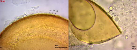
Outer layer (L1)- It is a hyaline, mucilaginous, often seen as a crushed fragile layer in both PVLG and melzer's reagent (melzer's: PVLG- 1:1). Thickness of the outer layer varies from 4.22-4.83μm in mature spores. Middle layer (L2)- It is a thick, continuous layer, which appears brownish in PVLG and reddish-brown in melzer's reagent. In mature spores the thickness varies from 3.36 μm to 5.63μm. Inner layer (L3)- It appears as a continuous pale yellow coloured layer in both PVLG and melzer's reagent. This thin inner layer remains attached to the second layer. The thickness of the inner layer varies from 1.44-2.13μm. |
|